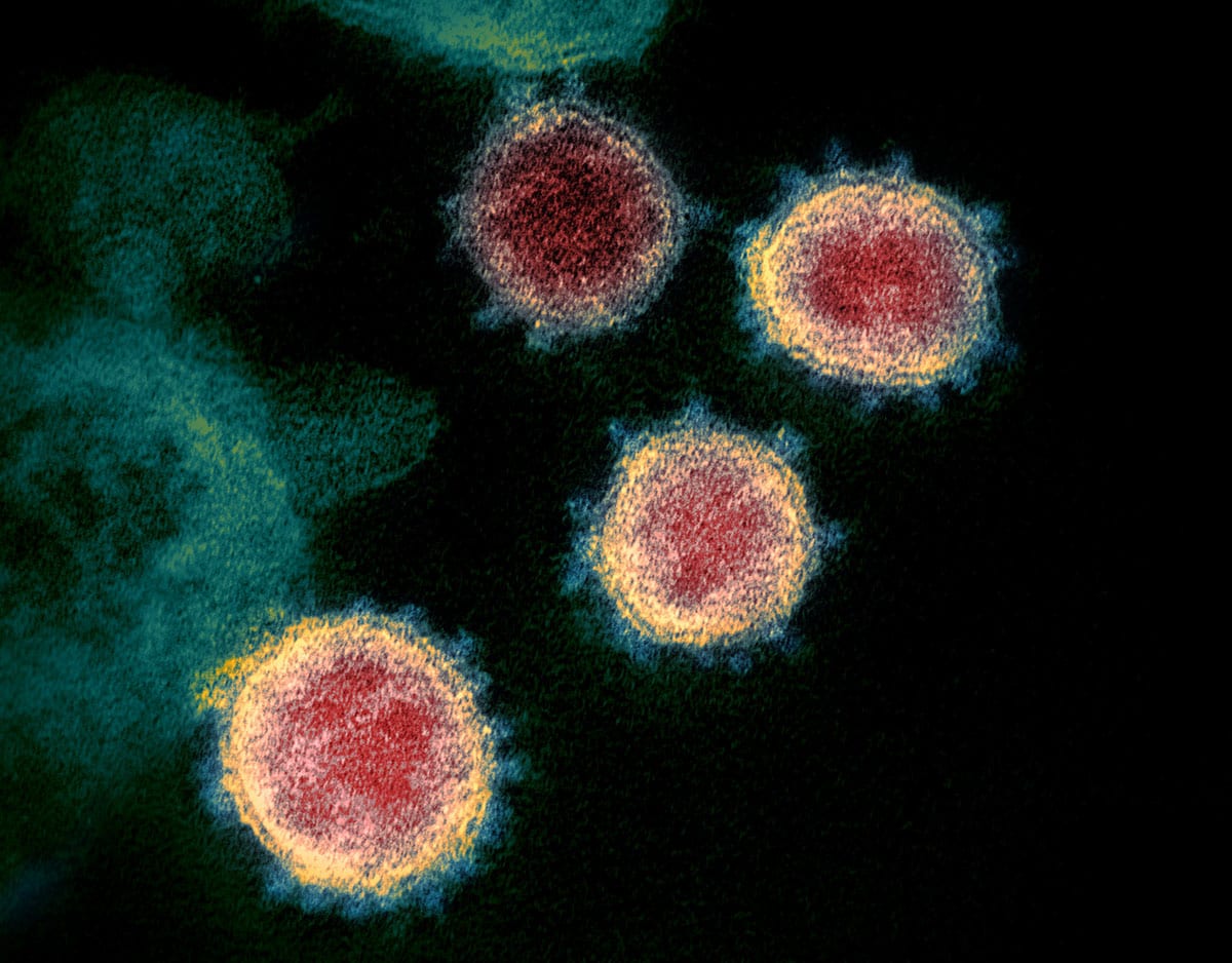
15 + Electron Microscope Image Of Covid 19 Virus HD Wallpapers. Virus particles are shown emerging from the surface of cells cultured in the lab. Taken by scanning electron microscope, images were colourised to better delineate virus from healthy cells.
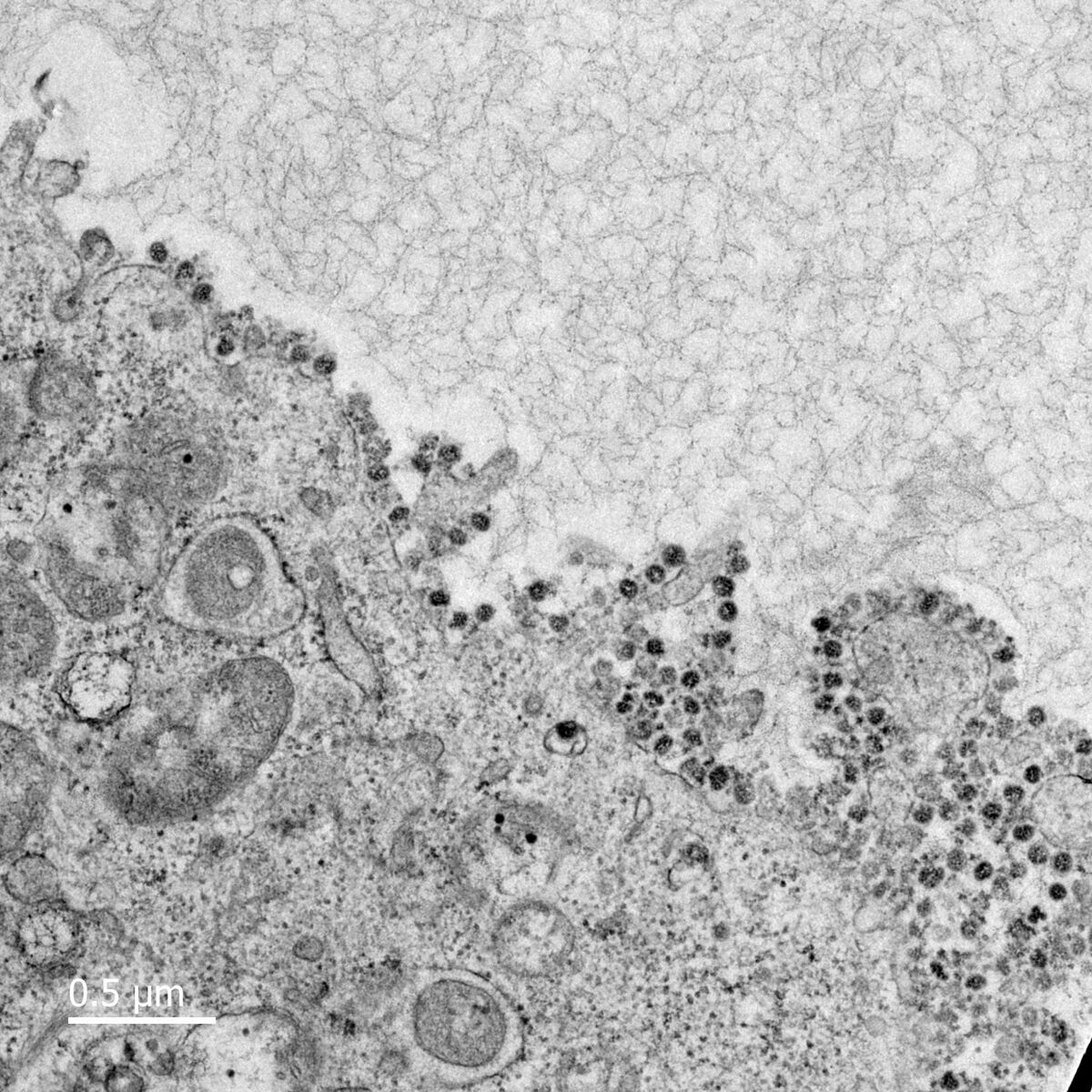
21 + Electron Microscope Image Of Covid 19 Virus Desktop Wallpaper
Download high resolution free stock photo collection online with photographer ad revenue sharing system at Freerangestock.com.
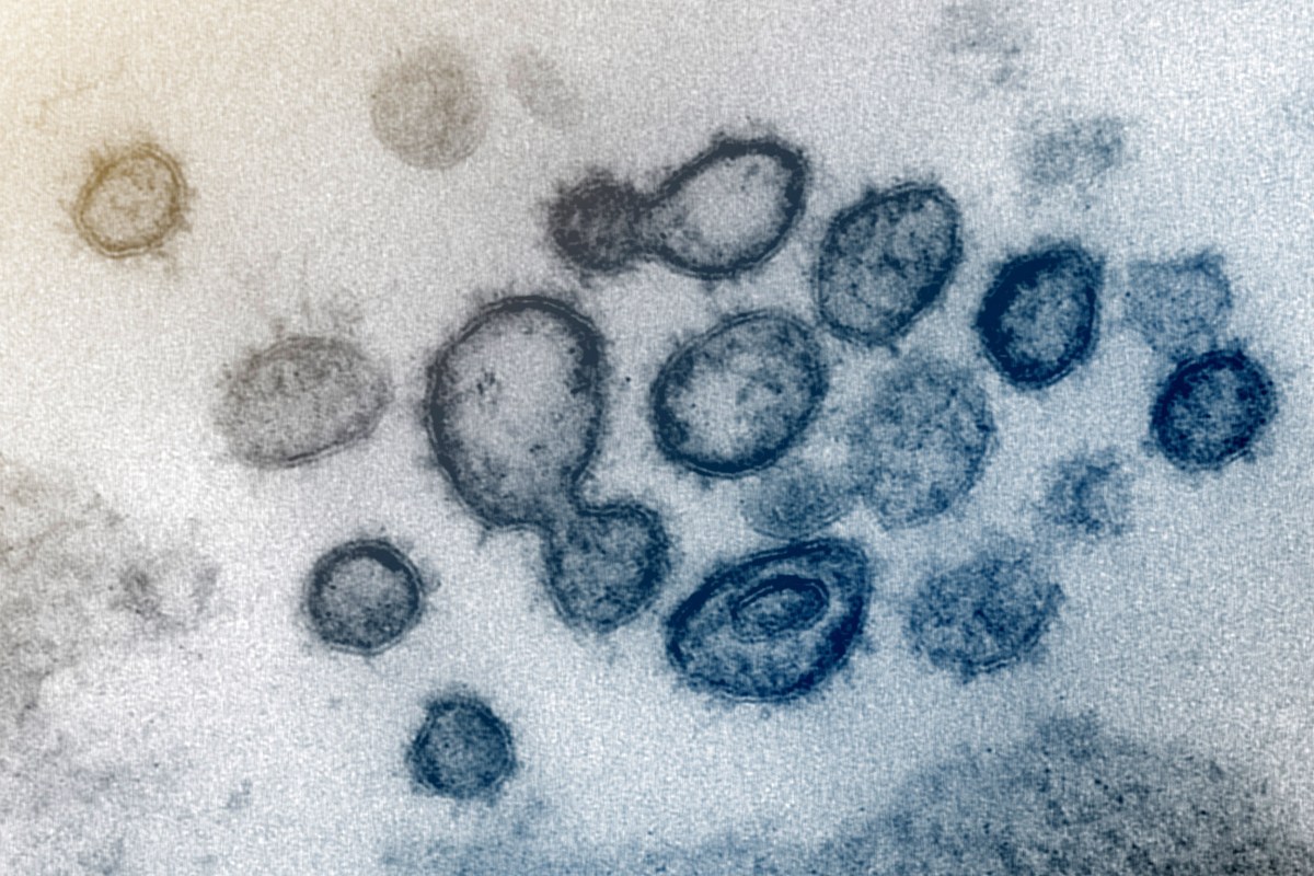
The why’s behind COVID-19 survival and immunity ...

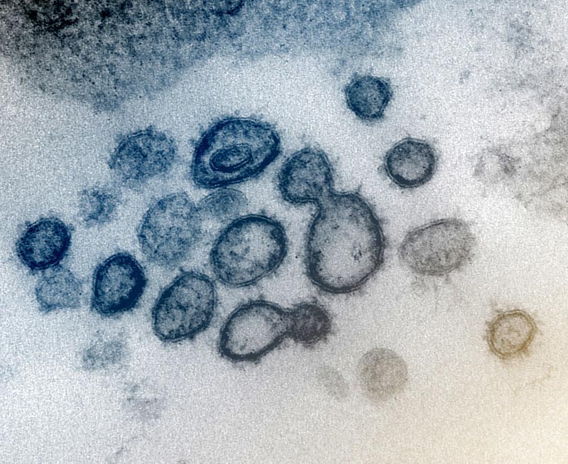
Figure. Electron microscopy images of thin sections and ...
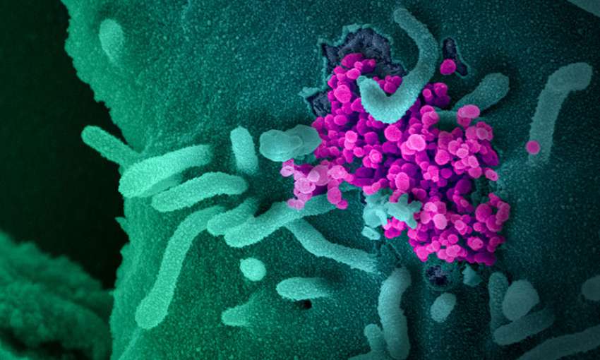
COVID-19 Expert Reality Check | Global Health NOW
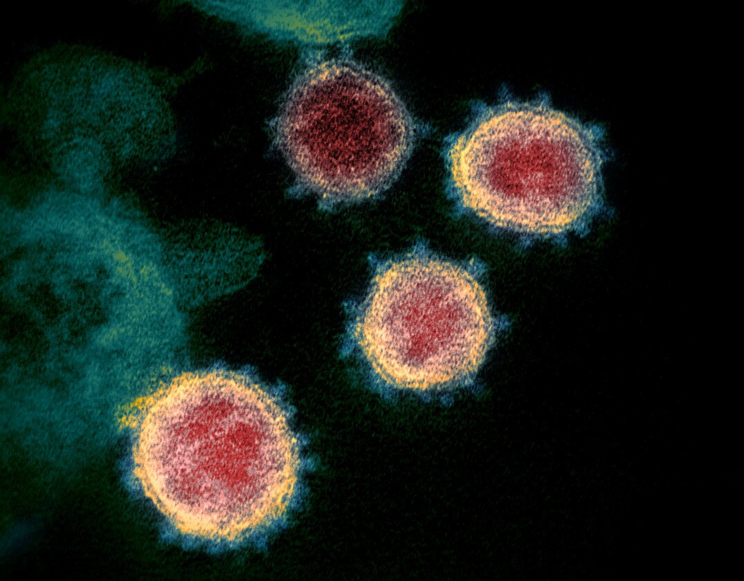
Study finds COVID-19 spread in China fueled by “stealth ...

Coronavirus Testing: Murphy Medical Associates Will Check ...
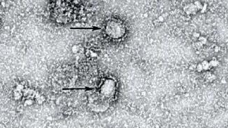
First image of new coronavirus released - CGTN

Total COVID-19 Cases In Georgia Rises To 11 | Georgia ...
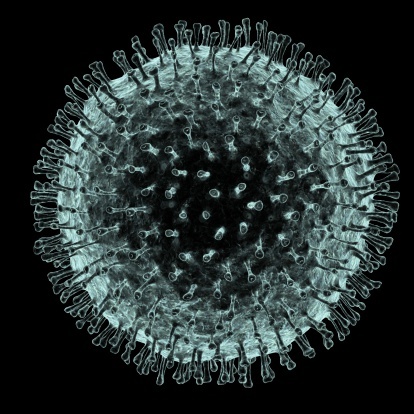
These 12 Viruses Look Beautiful Up Close But Would Kill ...

Coronavirus_SARS-CoV-2_Electron_microscope - Simmaron Research
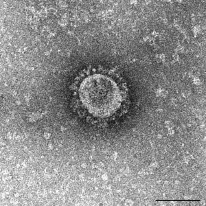
The MERS outbreak: an Asian perspective - On Biology
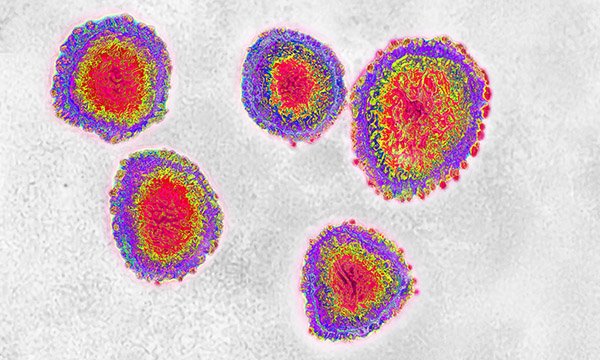
Transmission electron microscope view of coronavirus ...
We Must Protect the Frontline Warriors Against COVID-19 by ...

Pin on Really Small Things

MERS Coronavirus Particles | Colorized scanning electron ...
COVID-19: A Potent Reminder of the Challenge of Emerging ...
15 + Electron Microscope Image Of Covid 19 Virus High Quality ImagesResearchers at RML imaged samples of the virus and cells taken from a U. The virus was isolated from a. A Scanning Electron Microscope (SEM) scans the surface of a sample and records information that bounces back, similar to a satellite image.

