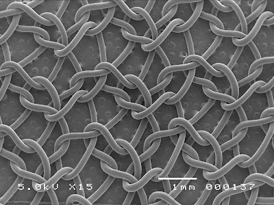
15 + Scanning Electron Microscope Images Of Cells Desktop Wallpaper. This study is the first applying Scanning Electron Microscope (SEM) to detect particle elements in urine. (Dr. To utilize these different SEMs, it is essential to recognize their features, as well as to understand the reasons for the contrast of SEM images.
21 + Scanning Electron Microscope Images Of Cells High Quality Images
Red blood cells (erythrocytes) are biconcave, disc-shaped cells that transport oxygen from.
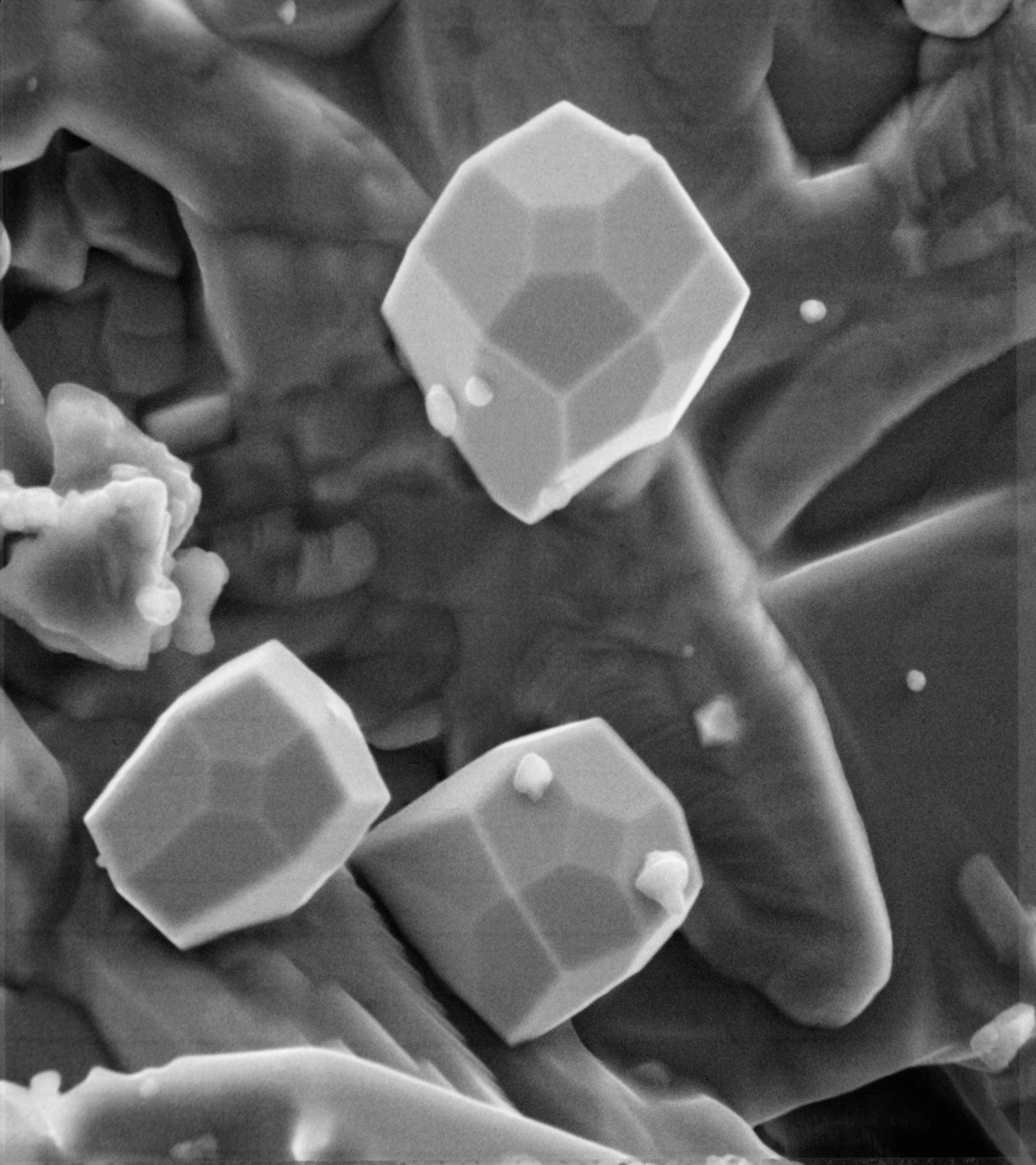
Scanning Electron Microscope View of Iron Crystal - Moon ...

Can people see eukaryotic cells under a scanning electron ...

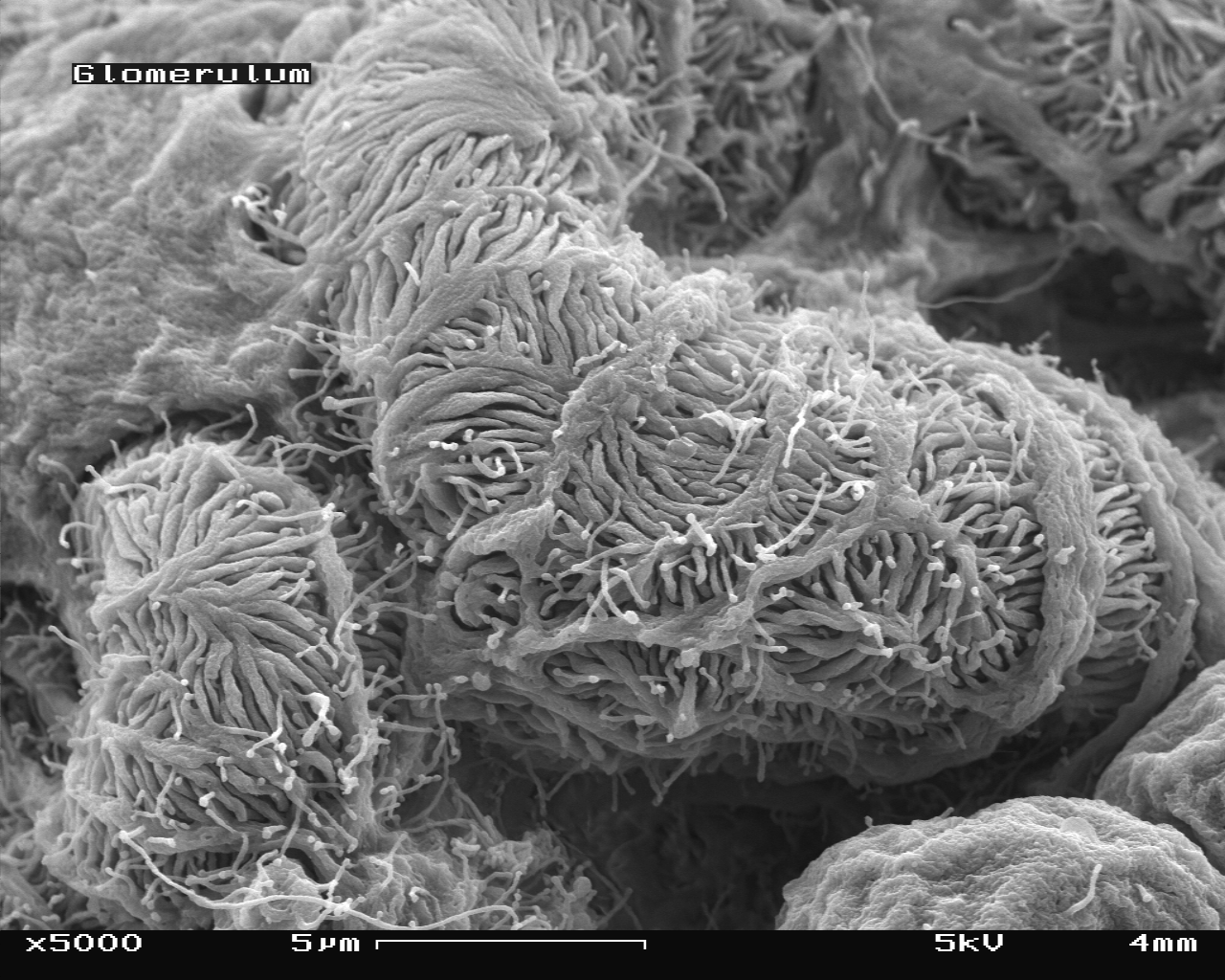
Glomerulus - wikidoc

Scanning Electron Microscope Of White Blood Cell High-Res ...

Scanning Electron Microscope Stock Pictures, Royalty-free ...
A scanning electron microscope image of a lymphocyte ...

scanning electron micrograph | NIH Director's Blog
Analysis, by scanning electron microscopy (SEM), of Vero ...

Pancreas Cell, Sem by Steve Gschmeissner | Microscopic ...

Plant Biology : Cell Press
Types of electron microscope — Science Learning Hub

Cell Culture | Cells in the SEM @ Cells Communicated

Untitled Document [ns.umich.edu]

Scanning electron microscope (SEM) image of a human white ...

Scanning Electron Microscope Of White Blood Cell High-Res ...
15 + Scanning Electron Microscope Images Of Cells High Quality ImagesNano Measurement and Characterization Tools: Scanning Electron Microscopy and Energy-Dispersive X-ray Spectroscopy. Image shows a cross-section of a cut leaf, itsupper epidermal layer, mesophyll layer with palisade cells and vascular bundles, and lower epidermal layer. SEM focuses on the sample's surface and its composition, so SEM shows only the morphology of samples.