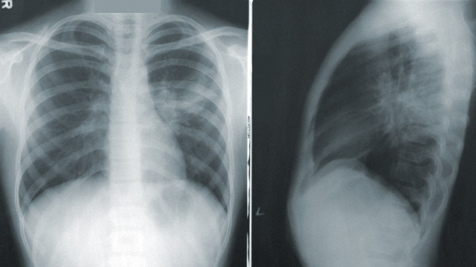
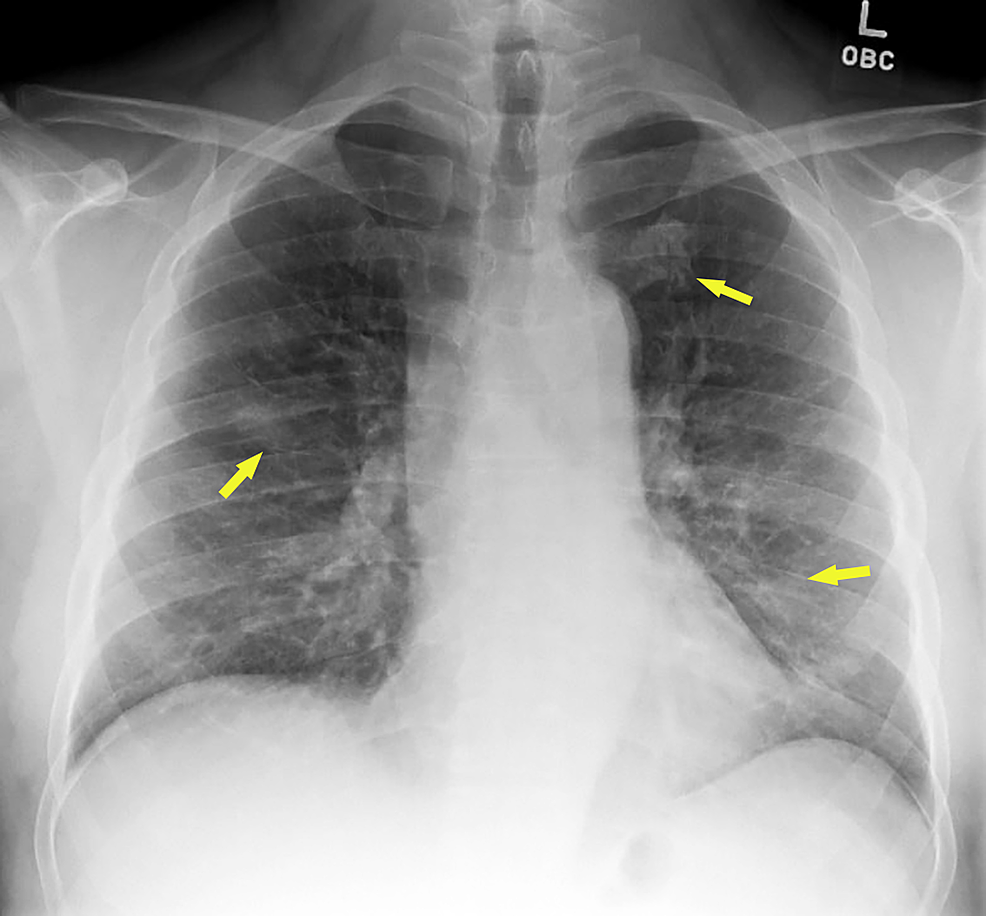
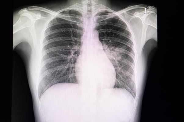
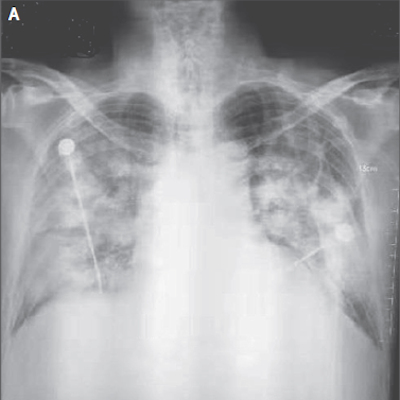
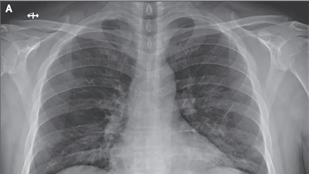
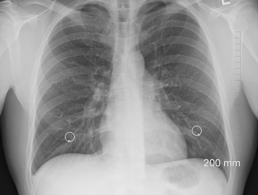
15 + Coronavirus X Ray Images Desktop Wallpaper. Medical imaging such as X-ray and computed tomography (CT) plays an essential role in the global fight. The images, released by the Radiological Society of North America, show what radiologists call ground glass opacity - the partial filling of air spaces.

21 + Coronavirus X Ray Images HD Resolutions
The coronavirus has brought the entire world to a standstill.
A neural network can help spot Covid-19 in chest x-rays ...


Behold.ai Partners with Apollo Hospitals Group for Rapid ...

MERS-CoV: Middle East respiratory syndrome corona virus ...

Coronavirus X Ray Of Lungs Seven Things That You Never ...
COVID-19 coronavirus compared to SARS outbreak: cases ...

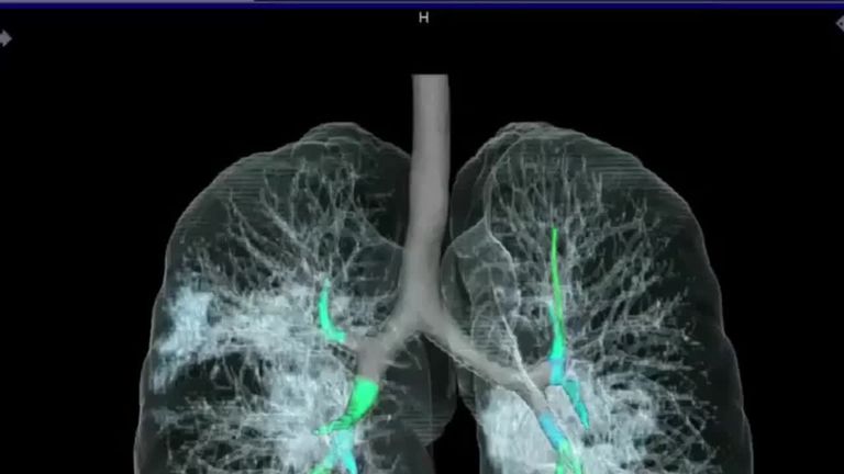
Coronavirus: What X-rays and CT scans reveal about how ...
Cureus | A Coronavirus Disease 2019 (COVID-19) Patient ...

front
New Research Finds Chest X-ray Not Reliable Diagnostic ...

Pediatric coronavirus disease (COVID-19) x-ray, CT in ...
X-ray, CT uncover novel coronavirus-infected pneumonia
Corona Virus Images Of Patients - Never lost your place

DarwinAI wants to help identify coronavirus in X-rays, but ...

MERS-CoV: Middle East respiratory syndrome corona virus ...
Dutch companies lead the way in developing x-ray software ...
15 + Coronavirus X Ray Images Background ImagesKeywords: Coronavirus; Pneumonia; Chest X-ray Radiographs; Convolutional Neural Network; Deep Transfer Learning. (I) Chest X-ray images have been used in the study. And now doctors are sharing scans of his lungs to reveal And when comparing image A to image F, it shows how the fluid in the spaces of the man's lungs became more pronounced over time. On the left we have positive (i.e., infected) X-ray images, whereas on the right we have negative samples.