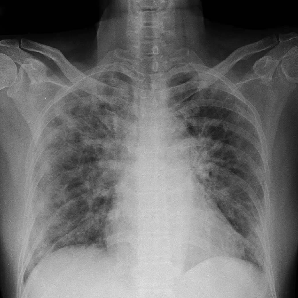
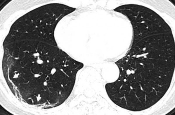
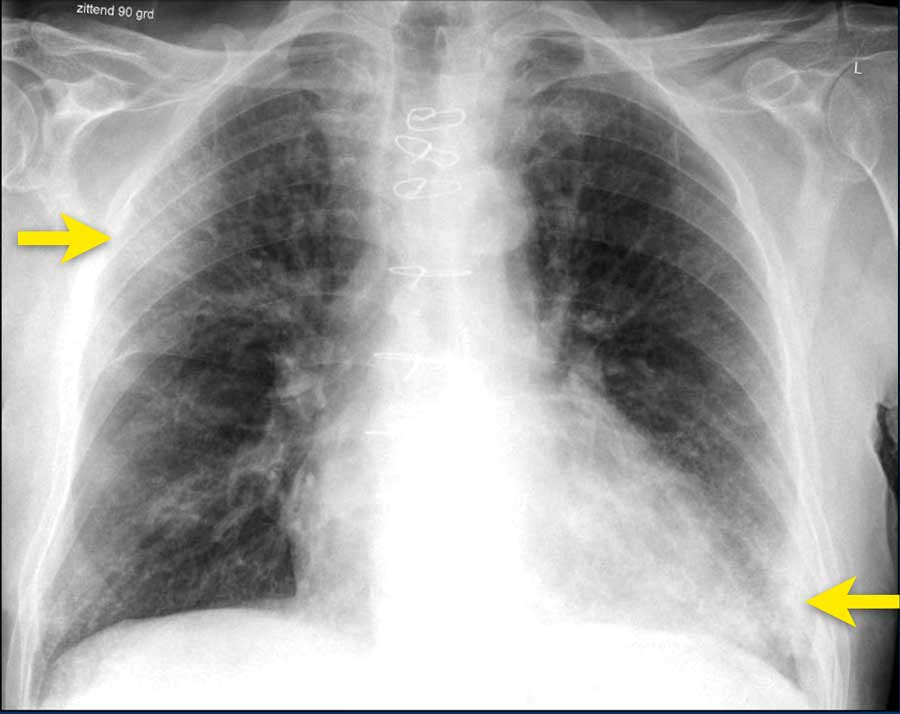
15 + Covid Lung Xray Picture HD Resolutions. LUNG SEGMENTATION NOTES: Lung segmentations in this dataset include most of the heart, revealing lung opacities behind the heart which may be relevant for assessing the severity of viral pneumonia. They are usually multifocal, bilateral and peripheral, but in the early phase of the disease the GGO may present as a unifocal lesion, most commonly located in the inferior lobe of the.

21 + Covid Lung Xray Picture Background Images
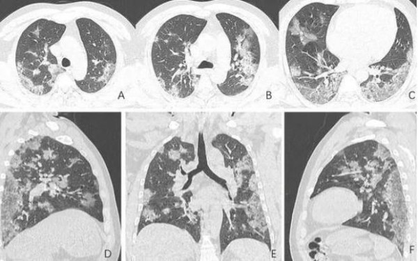
Upon analyzing CT scans and X-rays of the lungs of coronavirus patients, doctors have been able to identify common patterns and abnormalities, many of which are similar to those found in patients from the SARS and MERS outbreaks.

Covid 19 Lung Xray - covid 19 corona virus outbreak

AI tool gives doctors a new look at the lungs in treating ...

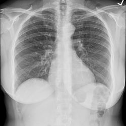
chest xray normal | Normal chest x-rays | Image ...
Coronavirus Update: Ravage Lung X-Ray Photos Show How ...
X-ray helps diagnose coronavirus in Vietnamese man

Coronavirus x-rays show terrifying damage in lungs of ...

Coronaviruses
Coronavirus Update: Ravage Lung X-Ray Photos Show How ...
The Radiology Assistant : COVID-19

Pin em x-ray

Chest x-ray showing upper respiratory tract infection and ...

Chilling coronavirus X-ray images show disease tearing ...

Clinical features and viral diagnosis of two cases of ...
Hidden in Plain Sight: The Prognostic Value of Chest X ...
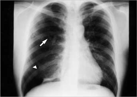
A posteroanterior chest radiograph reveals increased ra ...
15 + Covid Lung Xray Picture High Quality ImagesThey are usually multifocal, bilateral and peripheral, but in the early phase of the disease the GGO may present as a unifocal lesion, most commonly located in the inferior lobe of the. Use them in commercial designs under lifetime, perpetual & worldwide rights. Developed by Linda Wang and Alexander Wong at the University of Waterloo and IA firm..