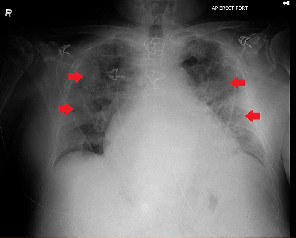
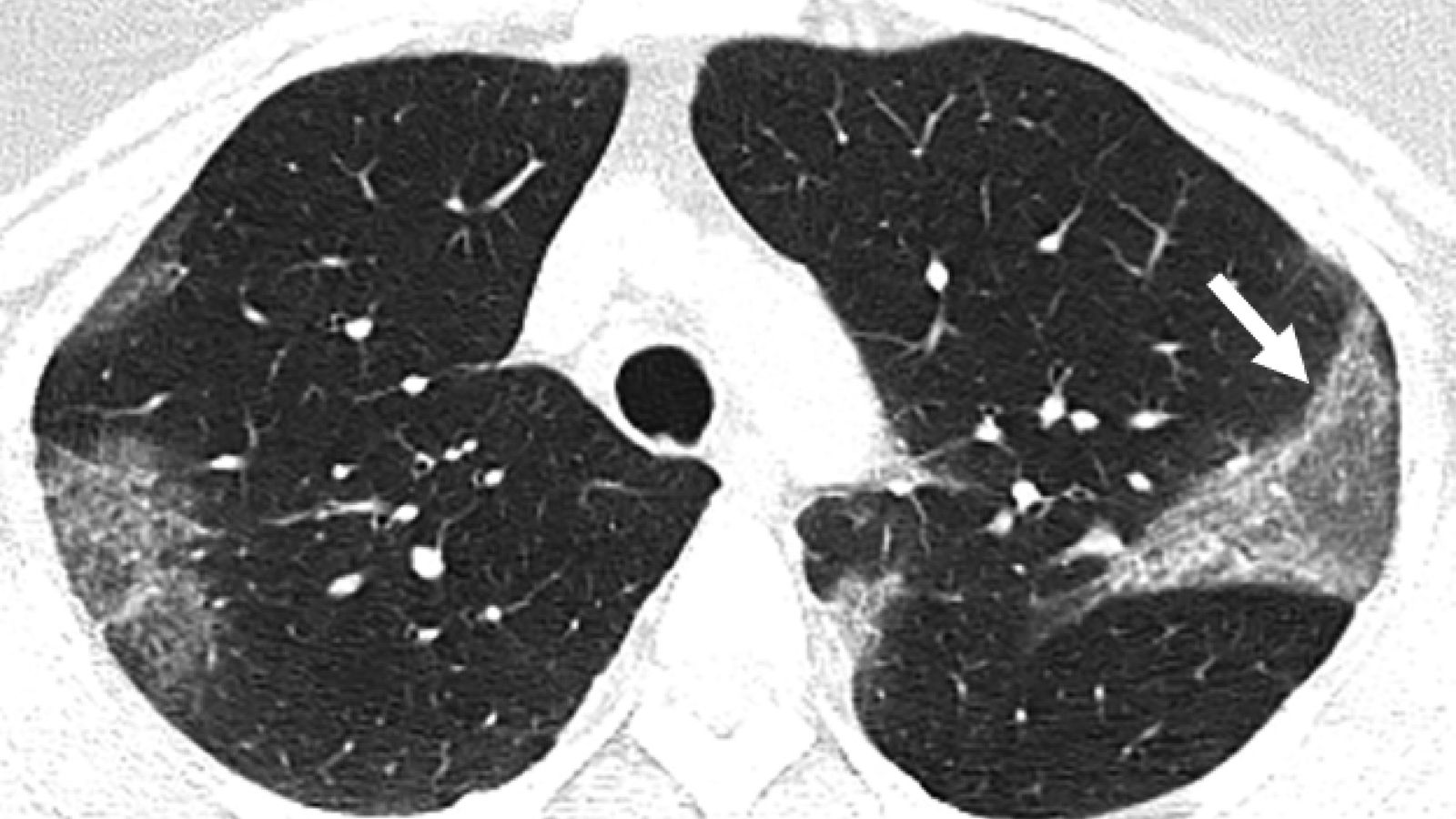
15 + Covid X Ray Image Vs Normal High Quality Images. I have seen in some analysis, people have combined the normal and pneumonia I plan to increase the robustness of my model with more X-ray scans so that the model is generalizable. Image: A large section of lung reveals what are called ground-glass opacities in the lungs.
21 + Covid X Ray Image Vs Normal Background Images
Medical professionals use X-rays to take pictures inside of X-rays were first discovered by Wilhelm Conrad Roentgen, a German physics professor.
Coronavirus: What X-rays and CT scans reveal about how ...


A Look At How The Immune System Fights The Coronavirus

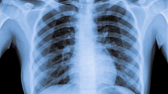
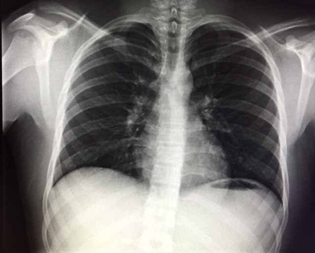
Normal chest x-ray | Image | Radiopaedia.org
The Radiology Assistant : COVID-19

Normal Chest X-ray Doesn't Rule Out COVID-19 | HappiDoc
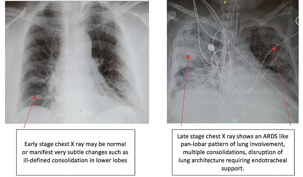
Cureus | Radiological Findings in Patients with COVID-19

A Beautiful Lung X-Ray! | trumpetlungs

Acute Respiratory Failure (ARF) | NCLEX Review & Nursing ...
Cureus | Coronavirus Disease 2019 (COVID-19) Complicated ...

Radiologists Describe Coronavirus CT Imaging Features ...
Coronavirus: What X-rays and CT scans reveal about how ...
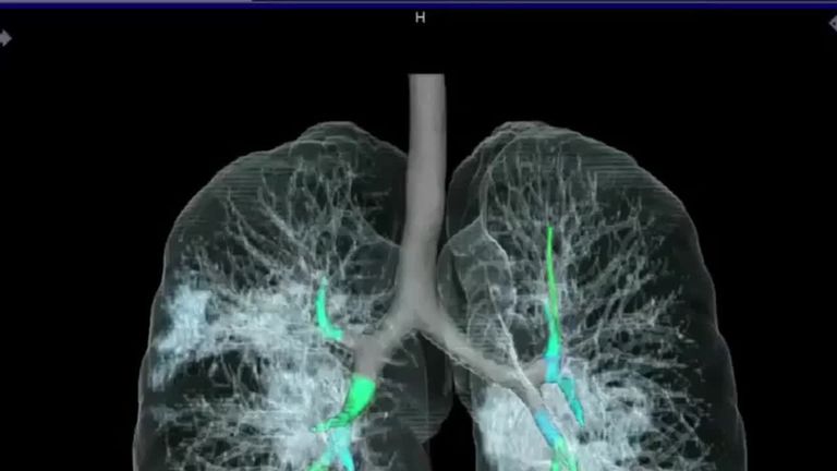
Digital Radiography (X-Ray) | River Radiology
X-ray helps diagnose coronavirus in Vietnamese man

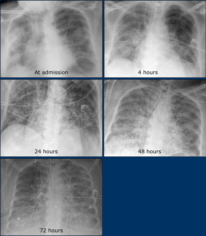
Chest X-ray frontal projection second day after admission ...

MIMIC Chest X-Ray database to provide researchers access ...
15 + Covid X Ray Image Vs Normal High Quality ImagesThe images from CT-scans and X-rays of patients with coronavirus show the extent of the damage to the lungs of the virus sufferers. Image: A large section of lung reveals what are called ground-glass opacities in the lungs. These medical images for his model have initially come out of the University of Montreal (Joseph Paul Cohen.